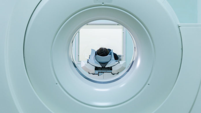
Medical imaging with CT, MRI, and PET can allow physicians and researchers to see inside a patient’s body, pinpointing disease and providing insight into physiological processes. These miracles of modern technology are incredibly complicated, with many sub-components working together and many settings available to adjust how the image is acquired.
While developing new imaging methods, scientists will use a stand-in for a human to image something without having to find a volunteer (or put one at risk). These devices are known as phantoms, and can be as simple as a large bottle of water. A graduate student running down to the local supermarket for a nice head-sized melon is a common sight around imaging labs.
Phantoms are also needed to calibrate imaging equipment, and to perform quality assurance tests. In these cases a more exacting stand-in is needed, and several manufacturers sell phantoms with well-defined chemical compositions and targets for imaging. More advanced imaging methods can require more capable phantoms to fully exploit the ability to make accurate measurements.
Advanced Phantom Engineering
Dr. Catherine Coolens of the Techna Institute has developed an advanced blood flow phantom that uses a pump and a system of tubes to accurately and consistently mimic blood flowing through tissue. It provides realistic flow conditions that can be applied to compare different scanners and imaging protocols, and even different modalities.
To help researchers understand the dynamics of this system, Dr. Coolens developed a mathematical model that describes the transport of fluid through the phantom and through the body. She also developed a second static – phantom to simplify the calibration requirements of scanners using IV contrast (licensed to Modus Medical Devices) and an open-source software tool for analyzing the images called the DCE QA Tool.
Together, these developments create a way to accurately calibrate and validate MRIs and CTs by ensuring that images taken using different scanners at various institutions remain consistent over time.
Knowledge Translation-as-a-Service
Bringing the development of a new phantom to the world imaging community through a Canadian company is already a big step from research to reality, but it can still take some time for imagers to find out about the innovation, order their phantom, and learn how to best apply it to their institution’s work.
To help accelerate its adoption, Dr. Coolens works with the Quantitative Imaging for Personalized Cancer Medicine (QIPCM) group at Techna. QIPCM provides services to help manage the advanced imaging needed for clinical trials in cancer. The first step is ensuring that the medical images for the trial will be high-quality and that there is consistency between all of the sites involved in a trial. QIPCM will help establish a common imaging protocol at each hospital participating in the trial, and perform quality assurance testing of the imaging equipment with phantoms such as the ones developed by Dr. Coolens.
To further expand the global reach of the phantoms, Dr. Coolens is working with collaborative networks such as the Quantitative Imaging Biomarkers Alliance and the NCI Quantitative Imaging Network, both of which aim to improve the use of new imaging methods in clinical trials and in clinical practice.
Providing services in close collaboration with experts like Dr. Coolens helps rapidly move advanced imaging methods—such as DCE imaging to measure blood flow in tumours—into practice. This model lets researchers see the impact of their physics innovations more quickly, while simultaneously helping clinical trials make use of cutting-edge techniques as soon as they are available. Working directly with the experts who developed and refined new techniques also helps cut out the learning curve needed to apply a method to practice.


