When medical conditions cause vision loss, that doesn’t necessarily mean that what a person sees goes dark. People with glaucoma or age-related macular degeneration can also have a secondary condition called Charles Bonnet Syndrome, where the gaps in their vision are filled in with things that aren’t actually there — an experience that can be very unsettling.
Vision neuroscientist Jennifer Steeves studies how the brain processes the visual world, both in health and in diseases like Charles Bonnet Syndrome. Her goal is to help uncover ways to treat patients with vision loss or brain damage.
“In my laboratory we study how the brain processes the visual world around us, how we recognize objects and scenes and faces,” says Steeves, professor of biology and researcher at York University’s Vision: Science to Applications (VISTA) program.
“Mapping the parts of the brain that process these different visual image categories, we have done work with patients who have specific brain damage.”
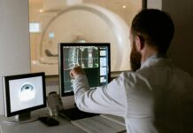
Steeves uses a non-invasive method called transcranial magnetic stimulation to apply neural noise to the brains of volunteers. It momentarily disrupts brain activity in specific regions that process visual information, allowing her to understand how each contributes to visual processing.
On the flip side, the same work illuminates how damage to certain regions of visual processing may contribute to conditions associated with vision loss, like Charles Bonnet Syndrome.
“Charles Bonnet Syndrome happens secondary to age-related macular degeneration or glaucoma, where individuals are losing their vision but they’re hallucinating visual images,” Steeves explains.
“Often people see things like textures or faces or objects that aren’t actually there. What’s actually happening is, there’s less input to the visual parts of the brain but the visual brain is still active, and so that’s creating these visual hallucinations. And so we’re hoping to bring these individuals into the lab to target those regions of hyperactivation and rebalance the activity in the visual cortex, so that they will no longer perceive these visual hallucinations.”
By mapping and quantifying brain changes and their effects, Steeves hopes her work will lead to treatment protocols across many conditions where there are imbalances compared to healthy brain activity. Disorders like depression and schizophrenia could also benefit from a similar approach.
With this map in hand, researchers will be better equipped to find the right direction.


