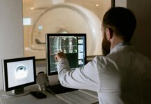
A specialized microscope could allow a dermatologist to pinpoint abnormal lesions and treat them at the same time, all without cutting the skin.
This new technique was developed by Haishan Zeng and Harvey Lui, professors of dermatology at the University of British Columbia and researchers at the BC Cancer Agency. At first, their goal was only to diagnose conditions like skin cancer, but then they wondered whether the same tool could be used to treat abnormalities by cranking up the power of the microscope’s laser.
The result is an instant diagnostic tool that could also offer same-day precision treatment by selectively closing the blood vessels that feed an abnormality. The study was published in Science Advances.
Laser therapy has already been widely used in medicine to treat excess blood vessel growth, which happens in many health conditions including cancers, port wine stains, and eye diseases like macular degeneration and diabetic retinopathy. But regular lasers are not precise because they rely on coloured molecules for selectivity, such as the red hemoglobin in blood, meaning that any blood vessel in the path of the laser could be destroyed — including ones that doctors want to avoid.
The new specialized microscope uses a femtosecond multiphoton laser, firing focussed pulses of light that last only quadrillionths of a second. Because the target will only absorb enough laser light if it is hit by two photons at the same time, areas outside the focal point are not affected. This gives incredible precision in every direction, including depth, both at the time of imaging and at the time of treatment.
At higher power, the multiphoton laser can selectively close single blood vessels based on location without harming their neighbours — even if those neighbours are closer to the skin’s surface. The authors validated their method by selectively closing capillaries and venules in the ears of mice.
While the technology has yet to be tested in humans, the laser can reach depths of up to about half a millimetre below the skin’s surface and about a millimeter below the brain’s surface. And because a laser beam can easily penetrate through the optically clear parts of the eye, it may even be used to non-invasively treat blood vessels supplying the retina at the back of the eye.
“We are not only the first to achieve fast video-rate imaging that enables clinical applications, but also the first to develop this technology for therapeutic uses,” said Zeng in a statement.
Because the microscope works on living tissue, it eliminates the need to cut out a sample of a skin lesion to send to the lab for testing, which can cause scarring and add weeks to a patient’s diagnosis. The team hopes to expand the applications for his tool by developing a miniature version that could be attached to an endoscope, allowing it to be used in the digestive tract.
Technologies like these that can give faster answers and non-invasive treatment to patients reduce anxiety and provide better care. Those are results that go much further than skin deep.


