Do you remember the first time you looked into a microscope? Maybe you were studying the structure of a snowflake, or the unseen organisms living in a scoop of pond water. The light microscope combines several lenses to magnify samples to allow users to see things that are normally too small to be seen with the human eye. This has made it one of the most powerful tools for studying cell biology.
Since its invention in the 17th century, the design of the light microscope has been further refined to see smaller and smaller details. However, the size of the wavelength of light itself seemed to limit light microscopes from resolving features smaller than about 0.2 microns, putting many important cellular features out of their reach. This is called the diffraction barrier.
To address this limitation, super-resolution microscopy (SRM) was developed to enable the study of sub-cellular processes in living cells using optical tricks to overcome the diffraction barrier. For the first time, this made light microscopes into nanoscopes.
Achieving greater resolution is particularly important in neurobiology, where observing living neurons allows us to understand how they interact, respond, and change over time. Prof. Yves De Koninck, Professor of Psychiatry & Neuroscience at Laval University, exploits this technology to study features of neurons that have never been seen before in live cells.
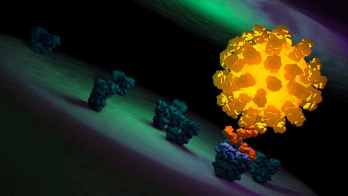
The power of SRM was validated in 2014 with a Nobel Prize in Chemistry for its inventors, but it remains difficult to understand. This animation by biomedical communicator Andrew Q. Tran visually explains key principles of several important SRM techniques, including foundational concepts of fluorescence and laser scanning microscopy.
Learn about how super-resolution microscopy works
Animation courtesy of Andrew Q. Tran
This technology allows researchers to understand live cell biology down to a new level, giving clues on how cells operate in both health and disease.
All images and animation under copyright © 2014 by Andrew Q. Tran, all rights reserved. Used with permission. To license, please visit andrewqtran.com.


