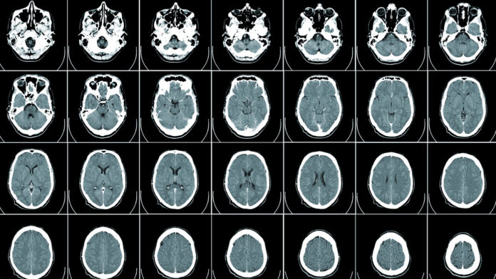
Canadian researchers made a surprise discovery when they examined human brain tissue from donors with multiple sclerosis (MS) and compared it to brain tissue from healthy donors: the brains of people with MS have unexpectedly high levels of a protein called calnexin.
Even more surprising, the researchers found that mice that lacked calnexin completely were also resistant to a mouse model of MS.
The authors of the new study, collaborators from the University of Alberta and McGill University, say that understanding the connection between calnexin and MS could open up new strategies for fighting disease progression.
MS is an autoimmune disease where the body’s own immune system mistakenly attacks cells of the central nervous system, including the brain and spinal cord. Specifically, the fatty covering on nerve cells called myelin is destroyed. Myelin is like the electrical insulation that allows signals to travel through nerve fibres, and when that covering is damaged, increasing physical and cognitive disabilities can arise.
Calnexin is chaperone protein that assists with protein folding and transport, and also plays a direct role in immune responses.
The researchers took mice that lacked calnexin and tried to induce experimental autoimmune encephalomyelitis (EAE), the most common mouse model of MS. EAE simulates the key features of MS in mice, including inflammation and loss of myelin.
The study found that mice that lacked calnexin were completely resistant to EAE induction, suggesting that they are also resistant to MS.
The calnexin-deficient mice showed no evidence of increased immune cell infiltration into the brain or spinal cord, and no increase in inflammation markers. Instead, they saw that the loss of calnexin drives a defect in the blood-brain barrier that reduces the passage of T-cells, a type of immune cell.
“It turns out that calnexin is somehow involved in controlling the function of the blood-brain barrier,” said co-author Marek Michalak, professor of biochemistry at the University of Alberta. “This structure usually acts like a wall and restricts the passage of cells and substances from the blood into the brain. When there is too much calnexin, this wall gives angry T cells access to the brain, where they destroy myelin.”
In other words, therapies targeting the unusually high levels of calnexin found in the brains of MS patients could help control the cascade of events that lead to the destruction of myelin.
“Our challenge now is to tease out exactly how this protein works in the cells involved in making up the blood-brain barrier,” adds co-author Luis Agellon, professor of human nutrition at McGill University. “If we knew exactly what calnexin does in this process, then we could find a way to manipulate its function to promote resistance for developing MS.”


