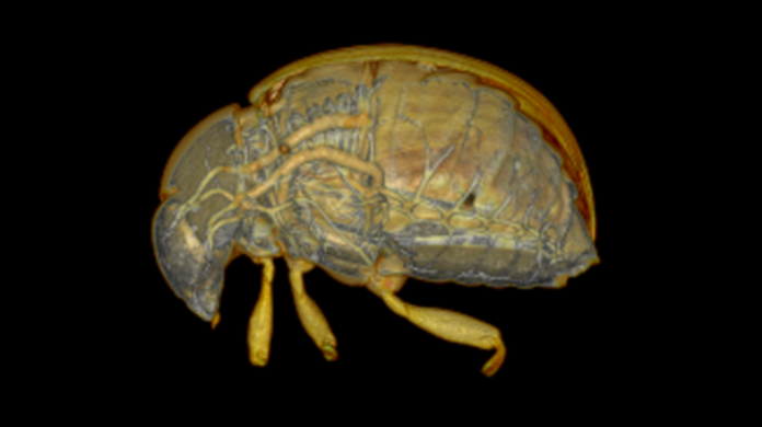
Researchers at Western University have just found a way to show you more insect than you’ve probably ever wanted to see.
The new technique, published last week in BioMed Central, can provide spectacularly detailed images of a living insect’s insides. The key: knocking the insects out with carbon dioxide before imaging.
Stay still
Insects have previously been imaged using X-ray micro-computed tomography (micro-CT), but the resulting images are usually blurry because the insects tend to move around during the scan. The alternative – killing the insects first – results in better images, but only provides static snapshots in time.
That’s not what Joanna Konopka wanted. The PhD candidate in Jeremy McNeil’s lab in the Department of Biology, wanted to study the internal development of the same insect over its full life cycle. To achieve this goal, she partnered with Danny Poinapen, a biophysicist at the micro-CT lab at Robarts Research Institute.
Poinapen knew imaging, but knew nothing about insects, Konopka was an insect expert, and together, they formed the insect imaging dream team.
Taking advantage of insects’ unique ability to withstand low oxygen environments, Poinapen and Konopka decided to try putting the insects into a state of suspended animation using carbon dioxide. The result were breathtaking, detailed images of an insect’s insides, down to a 20 micron (20 thousandths of a meter) resolution. The organs, reproductive system, and airways are clearly visible.
If you look at these pictures, you may never look at an insect the same way again. I, for one, never anticipated such complexity underneath an insect’s shell.
But the best part for science? There was no significant effect on the insect’s life history.
Get ready for your close-up
Using the technique, Konopka and Poinapen got high resolution images of the interior of adult Colorado potato beetles and true armyworms, but they expect it can be expanded to study the different development stages of these bugs. It is also detailed enough to determine mating status by looking for the presence or absence of a spermatophore – a capsule of sperm transferred from the male to the female.
Overall, this technique could have profound impact on the study of insects.


