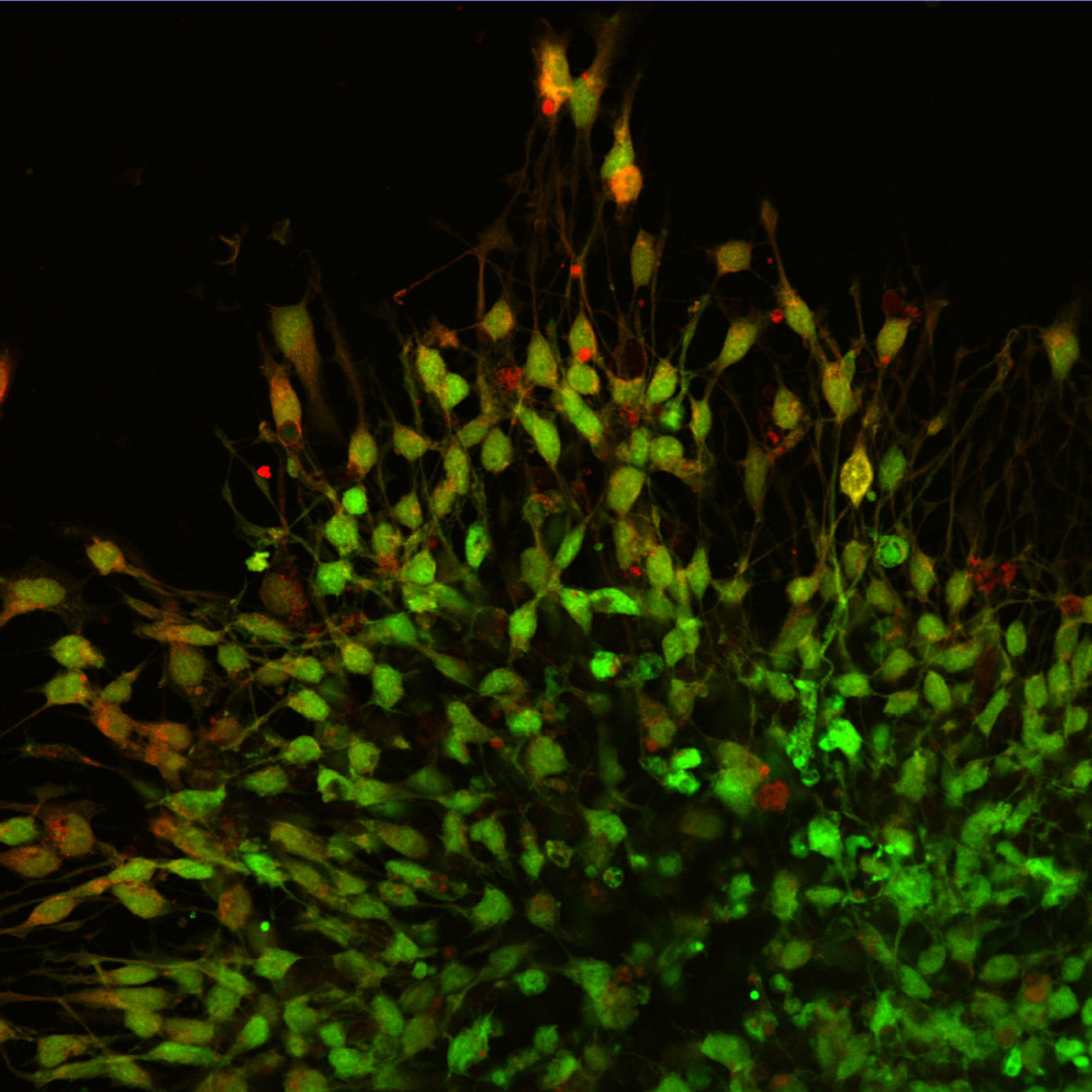
Walking into the Inner Spaces exhibit, my first thought was that this is the exact feeling of looking at cells through a fluorescent microscope. Tucked inside a darkened alcove, brightly coloured and larger-than-life images are lit from behind, making them appear as though light is emanating from within.
This is precisely the feeling that the curators intended: they opened a window for the audience to share in the same moments of discovery that real scientists experience in their everyday work.
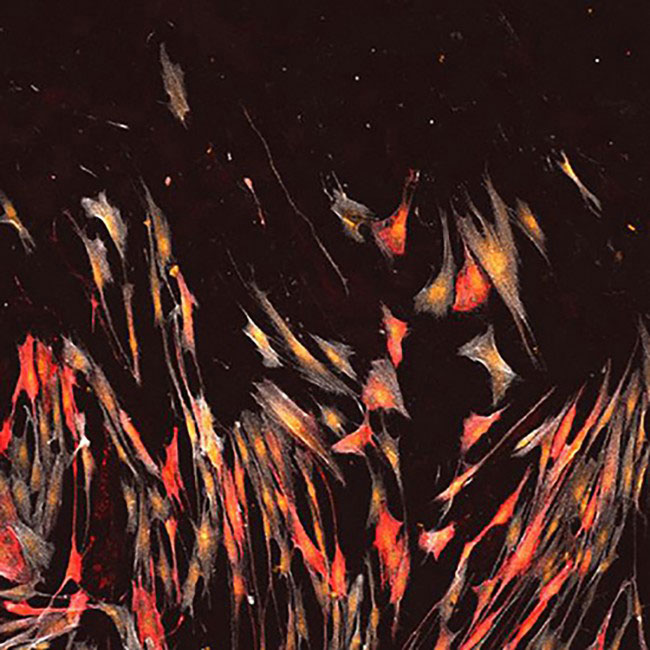
The images on display were not made to be works of art. They are real scientific images taken for real research projects. It just happens that biology, even on the microscopic level, is beautiful. And even if viewers don’t know exactly what it is they’re looking at, it’s this incidental beauty mixed with their curiosity that draws them in.
Curated by Kasey Ball, Anna Maria Kawecka, and Cassandra Kist, a trio of students from the University of Toronto’s museum studies program, Inner Spaces is now on display at the Ontario Science Centre until the end of May 2018 as part of the Scotiabank Contact Photography Festival.
The building blocks of science and inspiration
The Inner Spaces collection, a set of microscope images taken by scientists and engineers at the University of Toronto, balances aesthetics with diversity, using art to bridge a gap to new audiences and future scientists. The curators say that their request for images turned into a platform for telling stories, as there is so much behind the images that they wanted to communicate.
Many of the images focus their lens on human health and disease, and the potential treatments and cures under development in local laboratories.
Opening alongside science fair presentations by local youth via Visions of Science, Ball invited students to contemplate what the imagery meant to them.
“Now it’s your turn to discover Inner Spaces,” said Ball. “What I want you to do is to start from far away, and just enjoy what you see. Think about what the images look like to you. It will be different for everyone and that’s the best part. Then I want your curiosity to get the the better of you: come closer and find out what they are.”
Alexander Baker, a PhD candidate at the University of Toronto, contributed a piece called Bubbles. With bright blue spheres glowing against a black background, it’s one of the few pieces in the exhibit that has blurry edges. Like bubbles, Baker’s spheres have depth, and this shows in the image.
Shot using confocal microscopy, a technique that allows samples with depth to be imaged in thin slices, Baker later reconstructed Bubbles as a compressed z-stack, giving it a sense of dimension.
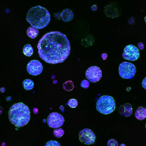
“Our cells are a bit like Lego bricks,” says Baker. “Those parts can only fit together and connect in certain ways, and when they do, they can build something big – like our eyes and brain, and even our entire bodies.”
Baker uses water-swollen hydrogels to help grow cells in three-dimensional clusters, helping them behave a bit more like they do in nature so that drugs can be screened more effectively in vitro, reducing the need to use animals in research.
“Being part of the exhibit and communicating this science to younger students made me think about the broader goals and broader picture of my research,” adds Baker.
“What other applications can we go after? How can I give an analogy that makes my work easier to relate to? How can I capture something beyond the message of the science and inspire people based on what they see?”
Never stop asking questions
Molly Shoichet, Ontario’s Chief Scientist and professor of chemical engineering and biomedical engineering at the University of Toronto, delivered the event’s keynote address.
“If you only remember one thing from today, I want it to be this: never stop asking questions,” said Shoichet. “If we stop asking questions, we’ll never discover something new.”
Shoichet recounted the story of George Washington’s death. In 1799, the year Washington died, antibiotics were not yet available. The standard of care was bloodletting: draining out “sick blood” from patients to help them recover. In just a few hours, over half of Washington’s blood had been withdrawn.
Unsurprisingly, when asked what they thought happened next, students called out, “he died.”
And they were right. The mix of blood loss and other complications killed him. Thankfully, medicine continued to evolve because people were driven to find better solutions to infectious diseases than bloodletting.
Many of the Inner Spaces images were contributed by researchers from Shoichet’s lab, depicting how stem cells can help replace cells damaged by disease or injury. None of these discoveries could have been made without asking questions about how things could be done differently to improve modern medicine.
In capturing and sharing these vibrant moments of discovery, Inner Spaces aims to inspire a culture of curiosity and wonder that leads to many more questions.